Microglia build and maintain the "roads" your brain needs to store memories
A protein called IL-33 keeps microglia working efficiently

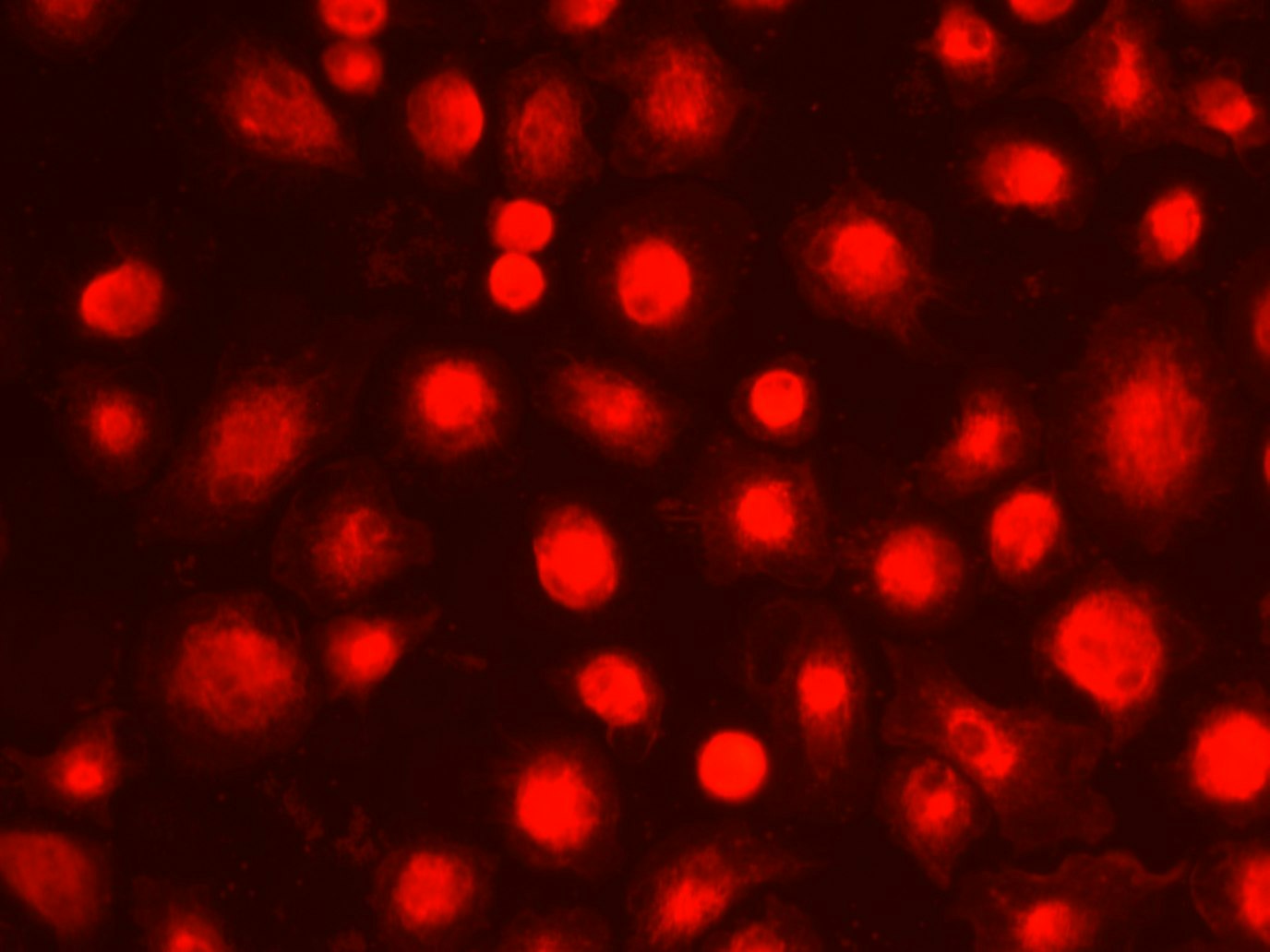
Jou63 via Wikimedia
What is memory? How can we improve it? How is it made? Understanding how memory forms on the molecular level will allow us to treat diseases such as Alzheimer’s disease and post-traumatic stress disorder, where memory formation and storage is impaired.
A study recently published in the journal Cell shows that the microglia in the area of our brain called the hippocampus play a key role in the creation of memories. They clear the surroundings of inhibitory proteins and eat old connections between neurons. Both these roles allowed new connections to grow and allowed memories to form.
Microglia are all-around heroes of the nervous system, taking on many different tasks, wearing many different hats. They protect the brain from pathogen invasion, remove overactive neurons, and more. Most importantly for our memory is their ability to clean. Cleaning is essential in the brain; a bit of leftover garbage – like misshaped or excess proteins – can lead to drastic consequences. One example of this garbage is aggrecan, a protein found in the space between cells, where it inhibits neuronal growth. When aggregan it isn't maintained and cleared regularly, its levels build up and stop neurons from growing.
Aggrecan and other brain garbage spew out a range of molecules that activate inflammatory genes in microglia, turning them into overactive cleaners who start removing too many of the connections between neurons. Instead of cleaners, microglia become demolishers, destroying the delicate balance of proteins in the environment that guide neurons to grow at the right speed and to the right areas. Eventually, if left unchecked, this rampant over-cleaning causes impaired neuronal activity and cell death.
Therefore, microglia behavior must be maintained at a proper level. This is accomplished in the brain in a number of ways, one being cytokine interleukin-33 (IL-33), a protein secreted by neurons. This new research shows that IL-33 keeps microglia in a healthy, cleaning state as long IL-33 is maintained at the right levels. This ensures that microglia eat the right amount of growth-halting proteins, such as aggrecan, in the cellular environment, and continue pruning back old neuronal connections to allow new ones to form.
Pruning neuronal connections is a critical memory-related function of microglia. Think of memory storage in your brain like a network of roads: if you have too many roads, you increase the chances of package out for delivery (a memory), of getting lost. If you have too few connections you might not have a road to get the package to its desired destination.
To show that IL-33 is important for memory formation, the researchers compared the activity of mice genetically engineered to lack IL-33 in their hippocampuses to the brains of normal mice. They tested the memories of the mice using a foot shock test. This involves the mice being shocked three times along with a stimulus such as white noise or bright lights. Then, they applied just the stimulus without the shock.
 and neurons (red)")
Microglia (green) and neurons (red)
GerryShaw on Wikimedia Commons
They predicted that a mouse with a healthy memory would remember the link between the stimulus and the shock, but a mouse without IL-33 would not. And they saw exactly this pattern: The mice without IL33 in their hippocampuses were much less able to link the foot shock to the sound and so in many cases did not react when they heard the sound. The normal mice scurried away after learning the link between shock and sound.
Next, they looked for exactly what IL-33 was doing in the brain. Comparing the hippocampuses of normal mice and IL-33 knockout mice, they found that a decrease in IL-33 levels lead to fewer connections between neurons in the hippocampus, making these mice less able to form new memories.
But how is IL-33 causing this change in the connections? The answer is through microglia.
The researchers found that injecting IL-33 into microglia led to changes in their activity. They became hungrier, and for specific things. One of the proteins they developed a taste for was aggrecan, the cellular space protein that stops neurons from growing. Using tracers specific for aggregan, they found microglia that had been injected with IL-33 were chock-full of aggrecan, and they saw a concurrent increase in neuronal connections. By eating aggrecans, the microglia removed roadblocks that allowed neurons to grow.
This appears to allow mice the ability to form new neurons. Without IL-33, neurons lost their connections and the mice lost their memories. In this experiment, they were unable to link a sound to a shock, but this could play out in any number of ways in the real world.
The brain is a complex organ and the role of IL-33 has not been fully explored – its levels naturally decrease with age, which may be related to age-associated memory loss, but high levels lead to inflammation in the brain. For this reason it's unclear if simply giving IL-33 as a therapeutic is a viable strategy. Other research has shown that microglia are modified through a range of different compounds, not just IL-33. IL-33 also has separate, unrelated functions in the immune system and is produced by immune cells. Could an infection also affect our memories? These questions and more regarding the role of microglia and IL-33 in memory will only be answered with more research.
Peer Commentary
Feedback and follow-up from other members of our community
Simon Spichak
Neuroscience
My own research focused on the way the immune system helps build and maintain the connections in the brain. I am particularly interested in how these signals help make complex memories. I’m sure there’s plenty of other proteins and signals we haven’t yet identified that help fine-tune our memories. This is a great breakdown of another exciting study in this field!
Minerva Contreras
Neuroscience
University of California, San Diego
As a glia enthusiast, I enjoyed reading this article. I have a question: when you say that microglia allow new neurons to be formed, do you mean new neuronal connections? Even though this study is in the hippocampus and neurogenesis is mostly explored there, I am wondering if this research directly addresses a connection between microglia and neurogenesis.