How does the developing brain learn to perceive the world before it can see it?
Our eyes work when we're born, even though they've never been exposed to light

Every day, within the belly of a towering concrete building at the University of California, Berkeley, researchers in the lab of neurobiologist Marla Feller strap on night vision goggles and get to work preparing paper-thin slices of retinal tissue in total darkness. The work is technically challenging, requiring surgical dissections and careful calibration of microscopes. These are non-trivial tasks even in a well-lit room, which these researchers must forego due to the light-sensitivity of their samples. They are driven by a central question: how do brains wire up effectively in order to allow an organism to perceive the world as soon as it’s born?
How the brain prepares itself for the requirements of independent life has been a central mystery in developmental neuroscience for decades. During embryo development, a hubbub of activity forges connections between neurons, which are later pruned, preserving only the most important ones. The refinement of these connections is a life-long process, but the bulk of the foundational work is done before maturity. Somehow, connections are being set up to perceive sensory information that the developing organism has yet to experience.
Feller got a hint about how brains could be primed to perceive light when she looked at the pattern of firing activity in the developing retina, a layer of neurons snuggled in the back of the eye. In 1994, as a new postdoc fresh from a PhD in physics, Feller was interested in applying novel imaging techniques to neuroscience.
"I barely knew what a cell was," she chuckled in an interview. Nonetheless, she was able to develop an imaging method to take movies of electrical activity across the entire retina. The first of her movies showed something surprising: even though there was no light to stimulate them, cells in the retina initiated waves of spontaneous activity.
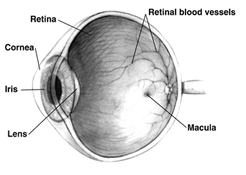
A diagram of the eye.
The waves were randomly distributed across the whole retina, radiating outwards from a central point in a starburst of neural activity, and they never overlapped with each other. In mice, they're first detected in newborn pups, who are born blind and with their eyes closed. Right around this time, critical connections are being made between neurons in the retina and the visual processing centers of the brain.
In the years since Feller's initial observations, scientists have found waves in the developing retinas of animals ranging from turtles to cats, leading them to posit that the spontaneous activity is a conserved mechanism by which developing visual systems organize themselves. In agreement with this hypothesis, when Feller's group manipulated the retina in 2016 to abolish the waves, they found that visual system neurons didn't wire up together properly.
The waves likely impact organization of the visual system through the effect they have on defining spatial relationships within the retina. As waves of neuronal firing sweep across the developing tissue, cells that are located close together tend to be activated together. These neighborhood interactions strengthen the connections between the cells close to each other, and to the neurons they connect to at higher levels in the brain, in a process called 'synaptic plasticity.' Neurons undergo changes that make them more responsive to signals from a cell that has fired on them many times. Waves prime neurons at higher levels of visual processing to be more responsive to retinal cells that are close together in space.
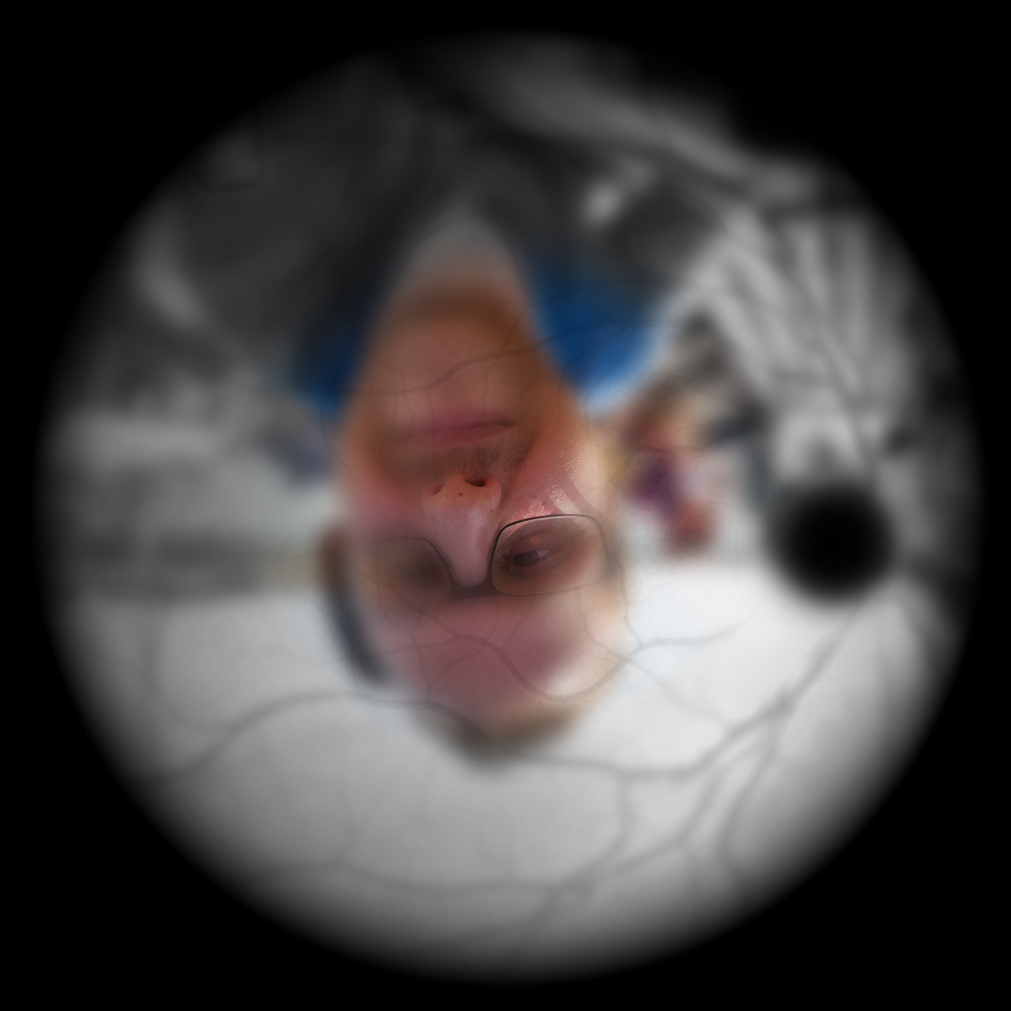
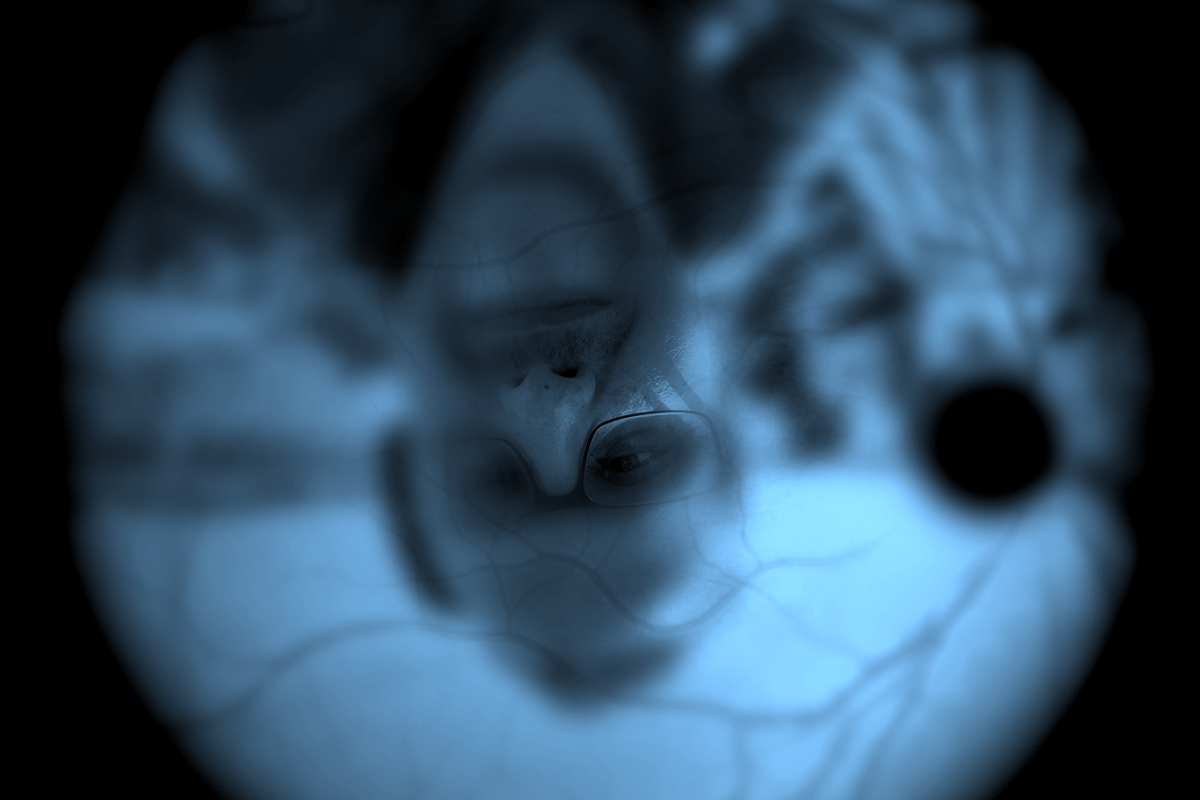
An illustration of what the retina might see, absent brain processing.
Strengthening the connections between neuronal neighbors is incredibly important in the development of the retina, because spatial arrangement of neurons forms the basis for high-level spatial perception. Cells in one spot of the retina are activated by light in one spot of the visual field; therefore, sloppy organization of spatial signaling relationships between neurons could result in a scrambled perception of 3D space.
There’s a lot left to explore – these days, Feller and her group are working to identify what exactly kickstarts the retinal waves, and to characterize which aspects are most important for later visual perception.
Research in this area is beneficial in multiple ways. The quality of retinal waves is impacted by a number of environmental factors, including alcohol and common prescription drugs. A good understanding of which chemicals diminish their activities can better inform guidelines given to pregnant women. This is a particularly poignant application to Feller, who while in the throes of premature labor with her son, realized that the medicines she was being given were similar to those she used to manipulate waves in the lab.
"I knew those drugs could cross the blood-brain barrier, and had to explain to my doctors the risk of the treatments," she said. Luckily, there are drugs available that do not cross the blood-brain barrier, and Feller's son is now a happy and healthy high school student. For Feller, the experience illustrates the power of research in informing safe therapeutic treatments.
Understanding the beginnings of neural processing is foundational to a complete understanding of the hierarchical organization of information processing in the brain. There’s a lot to be learned from how the massively impressive computers in our heads make sense of reality.
Peer Commentary
Feedback and follow-up from other members of our community
Benjamin Bell
Neuroscience
Johns Hopkins University
I've worked with retinal cells for years (ipRGC's), but still never knew about these waves. I wonder if the authors have compared retinal waves between animals with well-defined retinas (like the mice, turtles, and cats they speak of) to those with less specifically constructed retinas, but better understood cell lineages – such as the phototaxis neurons in C. elegans or the multi-retina of Drosophila? It seems like this might be an interesting way to root out the original function of the waves. Do they enable the animal to organize vision on 3D space, or is there a more basal, developmental function that would be seen in organisms whose only visionary requirements are "light" vs "no-light"?
Daniel Bear
Neuroscience
Stanford University
The waves have always been fascinating to me and important to my research, because they sit at the boundary of what we might call “innate” and what we think is “experience-dependent.” The nervous system doesn’t wire up properly without electrical activity like retinal waves, but the waves are sort of hardwired: it’s as if every animal had to watch the same set of movies, A Clockwork Orange-style, before being released into the world to have unique experiences. So it’s all part of this bigger question of how much is encoded in the genome vs picked up during a single lifetime. Where would you draw the line?
Lauren Mackenzie Reynolds
Neuroscience
McGill University
One thing that I find cool about this is that these retinal waves are happening with no sensory input, and are necessary for normal sight. But they also precede a critical period of experience: after the eyes open, they need to see things in order to properly organize the visual cortex. If an animal has one eye closed throughout the critical period, it will only ever see out of the eye that was open during the critical period, even if both eyes are open later in life. But not because the connection between retina and brain is impaired! They are still getting all of the information coming in through the eye, but their cortex has shuffled things around to give more processing space to their one eye that was open to experiences during the critical period. Nature and nurture: collaborators.